b value mri
Apparent diffusion coefficient ADC maps were created in the MRI scanner using a mono-exponential algorithm b value number of averages 0 1 10 1 25 1 50 1 100 1 250 1 450 1 1000 2 1500 3 and 2000 5 smm 2. With enhanced gradients the whole b ra in can b e scanned with in seconds.
A baseline b-value of 50 smm² is often used in liver diffusion-weighted imaging instead of b 0.

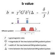
. Brain DW images obtained at b 3000 appear significantly different from those obtained at b 1000 reflecting expected loss of signal from all areas of brain in proportion to their ADC values. B γ² G² δ² Δδ3 Therefore a larger b value is achieved by increasing the gradient amplitude and duration and by widening the interval. DWI is done to determine the rate of molecular diffusion in different areas of the body.
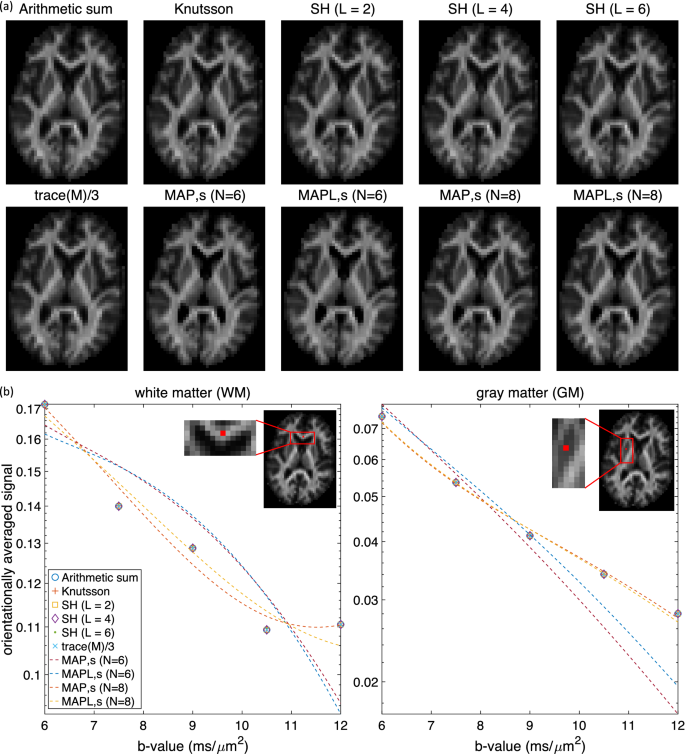
Pretreatment prediction of brain tumors response to radiation therapy using high b-value diffusion-weighted MRI. 2 article feature images from this case 4 public playlist include this case Promoted articles advertising. This ex vivo diffusion MRI data set consists of multiple b-values acquired within the same scanning session and since motion during scanning is absent the dataset allows voxel-to-voxel comparison across b-values directly on the acquired dataset.
The value of b was set at 0 30 50 80 100 150 200 400 600 800 smm 2 and varied throughout a single imaging session. Strength duration and the period b etween diffusion gradients. Multiple b values of 600 smm2 and higher are recommended to differentiate between benign and malignant.
B value measures the degree of diffusion weighting applied thereby indicating the amplitude G time of applied gradients δ and duration between the paired gradients Δ and is calculated as. Conventional MRI and multi- b -value DW images were collected before treatment at mid-stage evaluation and when conducting therapeutic efficiency assessment after chemotherapy. Code String 00189601 Diffusion b-matrix Sequence.
One hundred six patients underwent diagnostic multiparametric prostate MRI at 3T using an endorectal coil. MRI ADC Scans were obtained at six different B values. Calculated high b-value b1000 smm2and b2000 smm2 DWI were derived from the DWI dataset using DK and IVIM models.
In DWI the optimal b value is 600 smm2. Thirty-five PCa patients who were to be treated with radical prostatectomy underwent 3T DWI. Water diffusion MRIa potential new biomarker of response to cancer therapy.
The higher the value b the stronger the diffusion weighting. The best b-value combination was 0 and 600 smm2 and multiple b2. In contrast this molecular motion may be obstructed in certain pathological conditions - such as in the.
According to the Prostate Imaging Reporting and Data System version 2 high b -values 14002000 smm 2 are favored over standard b -values 8001000 smm 2 for improving tumor detection since they can qualitatively distinguish lesions and normal prostate tissue. The b value is used in MRI in the context of Diffusion Weighted Imaging DWI. Crossref Medline Google Scholar.
Studies have reported that the use of b values higher than 1000 smm 2 and 2000 smm 2 improves tumor localization and the contrast between benign and malignant lesions in the prostate and the breasts 12 14. The b -value is one of the primary parameters influencing DWI results. 104 Patterson DM Padhani AR Collins DJ.
Initial b-value - Questions and Answers in MRI b0 vs b50 In body imaging a starting b-value of 50 smm ² instead of b0 is often used. B 0 100 200 500 1000 2000 presented sequentially in a single stack with calculated ADC as a separate stack. In general approximately 1000 smm 2 is the maximal b value for DWI 5 11.
The lesionnormal parenchymal ADC ratio for b600 b1000 and multiple b2 better distinguished between benign and malignant lesions. Note how the T2 component is less pronounced with higher B values. In general in healthy tissue molecules of water and other chemicals are not stationary but moving about.
As demonstrated in Table 3 a high positive predictive value 953 indicates that DWI at b 3000 smm 2 is clinically useful to predict high-grade tumor hyperintense or markedly hyperintense on DWI or higher than score 4 though it is unlikely to replace biopsy to determine tumor grade and pathology. 2015 designed a study to correlate the accuracy of 3T MRI in which DWI occurred with a b-value of 2000smm 2. Double 00189147 Diffusion Anisotropy Type.
The degree of diffusion weight in g correlates with the strength of the diffusion gradients characterized b y the b - value which is a function of the gradient related parameters. Depending on the organ being imaged b-values typically range from 50-1000smm 2. Sequence 00189125 MR FOVGeometry Sequence.
Important characteristic of a thermistor B parameter B-factor in crystallography Constant b in GutenbergRichter law Acquisition parameter in diffusion MRI This disambiguation page lists articles associated with the title B-value. Facts about the dataset. Sequence 00189119 MR Averages Sequence.
B-value may refer to. Cardiovascular Imaging - Supplies - Open Directory Project - MRI Technician and Technologist Career - Abdominal Imaging - Online Books B-Value The b-value is a factor of diffusion weighted sequences. The b factor summarizes the influence of the gradients on the diffusion weighted images.
Sequence 00189152 MR Metabolite Map. Two recent studies explored DWI at an ultra high b-value of 2000smm 2. Sequence 00189118 Cardiac Synchronization Sequence.
5 b-value b 0 188 375 563 750 smm2 DWI and high b-value b0 1000 and 2000 smm2 DWI were acquired. Consequently when all other imaging parameters are held constant b 3000 DW images appear significa. High b-value diffusion-weighted MRI of normal brain.

Diffusion Weighted Imaging Radiology Reference Article Radiopaedia Org
Diffusion Weighted Imaging Radiology Reference Article Radiopaedia Org
Computing The Orientational Average Of Diffusion Weighted Mri Signals A Comparison Of Different Techniques Scientific Reports

Apparent Diffusion Coefficient Radiology Reference Article Radiopaedia Org
Frontiers Fixel Based Analysis Effectively Identifies White Matter Tract Degeneration In Huntington S Disease

Hyperpolarised 13c Mri Identifies The Emergence Of A Glycolytic Cell Population Within Intermediate Risk Human Prostate Cancer Nature Communications
Diffusion Weighted Imaging In Hemorrhage Radiology Key

Principles Of Diffusion Tensor Imaging And Its Applications To Basic Neuroscience Research Neuron

Mri Planes For Mri Head Scan A Axial B Coronal C Sagittal Mr Download Scientific Diagram

Comprehensive Mri Assessment In Acute Stroke Using Dwi Pwi And Mr Download Scientific Diagram

Signal Intensity Of Dwi And Adc In Diffusion Restriction Increased Download Scientific Diagram
Tensor Valued Diffusion Encoding For Diffusional Variance Decomposition Divide Technical Feasibility In Clinical Mri Systems Plos One

Conventional Mri Dwi And Adc Images Of Braf V600e Mutant And Download Scientific Diagram
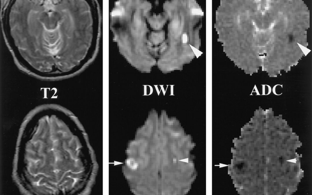
Diffusion Weighted Imaging Matlab Number One

Apparent Diffusion Coefficient Radiology Reference Article Radiopaedia Org

Principles Of Diffusion Tensor Imaging And Its Applications To Basic Neuroscience Research Neuron

Cerebral Micro Structural Changes In Covid 19 Patients An Mri Based 3 Month Follow Up Study Eclinicalmedicine
0 Response to "b value mri"
Post a Comment